Diagnosing Skin Cancer With a Biopsy
A biopsy is a diagnostic measure that involves a dermatologist taking a small piece of abnormal tissue in order to more closely inspect it. There are several different types of biopsies including excisional biopsy, punch biopsy, incisional biopsy, fine needle aspirate and shave biopsy. After removal, skin samples are placed in a solution and thin slices are put on slides to be viewed under a microscope.
The Use of Dermoscopy by Medical Dermatologists
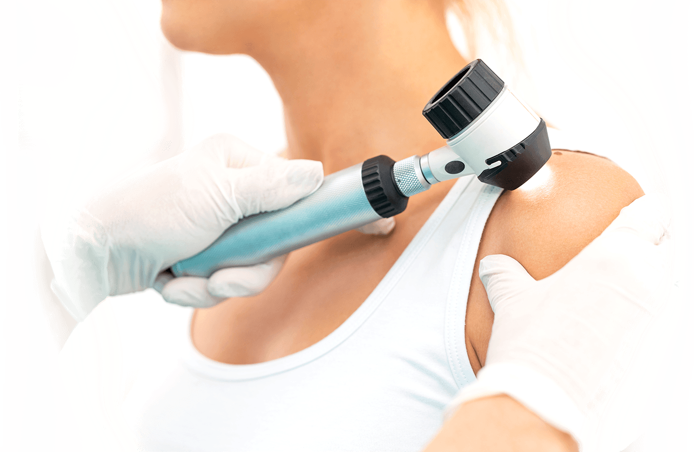
Dermoscopy is a method of examining skin abnormalities in order to differentiate between benign and malignant skin lesions. It is performed using a dermatoscope which has a high quality lens and light source. In order to cope with the reflection of light on the skin surface of the patient, physicians will use either a liquid immersion technique, which is using oil or liquid on the skin and lens and pressing the lens directly against the skin, or a polarized lens. This technique helps the physician get a better look at moles and skin lesions.
Mole Mapping for Skin Cancer Diagnosis
Mole mapping is a way to track changes in skin pigmentation and lesions in people who are at a high risk for melanoma. The method is used in hopes of catching a cancerous skin lesion in the earliest possible stages. The method generally used is to take whole body digital photographs and possibly close up pictures of areas of concern. The photographs are then examined by a skin cancer specialist.
To learn more about our skin cancer diagnosis methods, contact the dermatologists at any one of our various Orange County dermatology offices.


